Lateral Capsular Ligament Knee Mri
It is attached below to the posterior margin of the head of the tibia. Although the medial collateral ligament MCL is frequently injured descriptions of the appearance of the medial capsular and supporting structures of the knee at MR imaging are often not very detailed 1.
 Unstable Knee Complete Orthopedics Multiple Ny Locations
This has been referred to as the lateral capsular sign 1 which is best seen on the anteroposterior view of the knee.
Lateral capsular ligament knee mri. Low PPV findings on MRI include. It is also attached to the upper margin of the intercondyloid fossa and posterior surface of the femur close to the articular margins of the condylesIt crosses the popliteal fossa from medial to lateral. Investigators involved with the study of this structure have named it the anterior band of the lateral collateral ligament 1 and anterior oblique band 6.
However coronal MRI can demonstrate soft tissue or bony Segond avulsions with isolated posterolateral corner injury 2 9. Anterolateral stabilization is provided by the capsule and iliotibial tract. 2 Cooley VJ Larson RV Harrington RM.
The anterior oblique band AOB is seen on axial MR images as a thin hypointense band extending from the lateral collateral ligament to the lateral tibia and the iliotibial tract Fig. Positive predictive value PPV is low 9 medially 13 laterally 7. 13 The OPL connects the posterior medial and lateral knee attaching medially to the capsular arm of the POL and the SM and coursing superolaterally along the posterior joint capsule to the arcuate ligament and lateral head of the gastrocnemius.
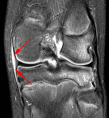
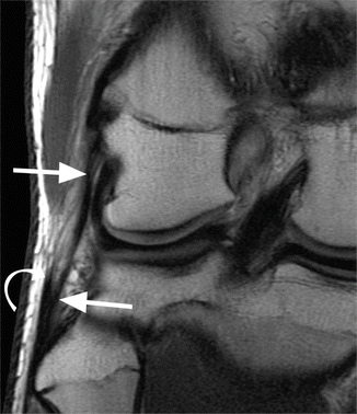
Chondral thinning and chondral contusions noted in the lateral compartment. Diagnosis typically takes place arthroscopically. Interposition of fluid between the meniscus and the medial collateral ligament 2.
According to one study had a PPV of 0 medially and 50 laterally 7. 2 Biceps femoris muscle. A mid-third lateral capsular ligament.
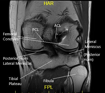
The knee is the joint most commonly examined at magnetic resonance MR imaging. They divided this structure into meniscotibial and meniscofemoral. The oblique popliteal ligament originates from the adductor tubercle of the medial side of the femur.
Described the mid third lateral capsular ligament as a thickening of the lateral capsule of the knee attaching to the femur in the region of the lateral epicondyle with capsular attachments to the lateral meniscus and insertion onto the tibia posterior to the Gerdy tubercle and anterior to the popliteal hiatus. Magnetic resonance imaging MRI is a radiologic procedure that uses a magnetic field and radio waves to develop detailed image cross-sections of the body including the knee1. Associated injuries include 13.
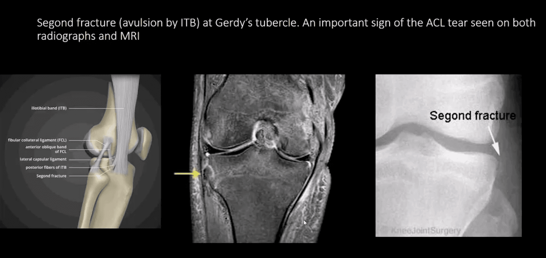
MRI is essential in all cases of Segond fractures to identify internal derangement. Grade 2 sprain of the tibial collateral ligament with partial thickness tear of the ligament itself proximally. A further 20 knee MRI studies with a Segond fracture were assessed to determine a relationship between the fracture and the anterolateral ligament.
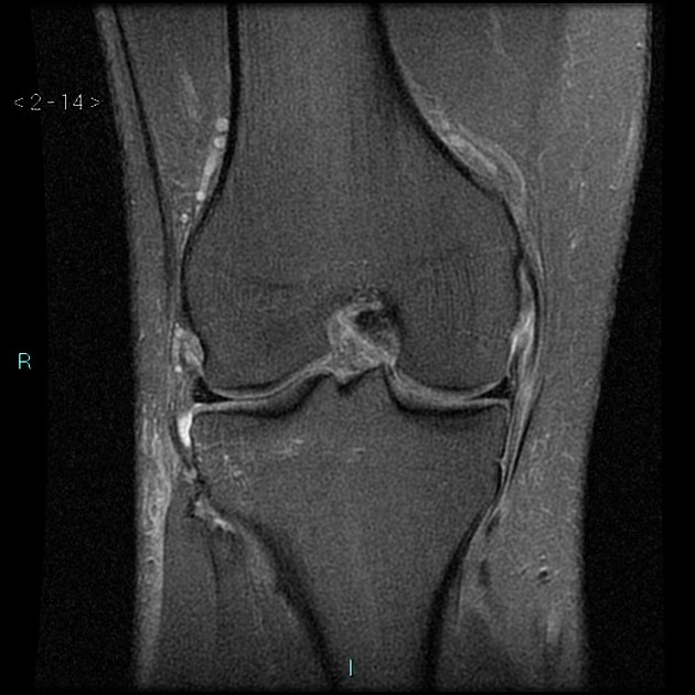
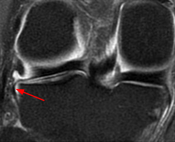
1 Vastus lateralis muscle. The posterior cruciate ligament PCL is one of the two cruciate ligaments that stabilize the knee joint. An avulsion fracture Segond fracture of the lateral tibia secondary to injury of the meniscotibial portion of the middle third capsular ligament is considered indirect evidence of an ACL injury Fig.
It provides dynamic stability to the knee during flexion causing increased tension in the joint capsule and POL which increases medial stability and in the OPL which assists with lateral capsular stability. In a 2000 review of the MRI appearance of structures that comprise the post-erolateral knee LaPrade et al. Small flap tears noted in the anterior and posterior horns of the lateral meniscus series 5 images 17 and 18.
Gross anatomy The PCL attaches to the posterior intercondylar area and passes anterosuperiorly to insert into the lateral surface of the. These structures can be demonstrated with routine spin-echo magnetic resonance MR imaging sequences performed in the sagittal coronal and axial planes. T2-weighted FATSAT Sagittal view.
In all 53 cases a structure was present along the lateral knee connecting the distal femur to the proximal tibia with meniscofemoral and meniscotibial components. Mid-Third Lateral Capsular Ligament Originates on the femur just anterior and proximal to the lateral epicondyle posteriorly to the supracondylar process of the lateral femoral condyle extends to the meniscus then further to attach to the tibia posterior to Gerdys tubercle at its anterior aspect and ends at the anterior edge of the popliteal hiatus. Meniscocapsular separation is usually diagnosed arthroscopically and the positive predictive value PPV of MRI has been traditionally described as being low 3 as low as 9 medially and 13 laterally.
Magnetic resonance imaging of the torn anterior lateral ligament ALL is inconsistent and subject to significant intra- and interobserver variability. Am J Sports Med 199927469-475. The effects of grade III posterolateral knee complex injuries on force in an anterior cruciate ligament reconstruction graft.
3 These structures of the PMC are. On MR images the lateral collateral ligament appears as a straight homogeneous hypointense structure on all sequences Fig. 7 reintroduced the anatomical.
Effect of lateral ligament reconstruction on intra-articular posterior cruciate ligament graft forces and knee motion. In 2013 Steven et al. The lateral aspect of the knee is stabilized by a complex arrangement of ligaments tendons and muscles.
Disruption of the ACL is the most common however there are additional frequently encountered injuries. Low predictive value MRI findings that have been correlated with meniscocapsular separation include 1-2. MRI of the knee.
 Pdf Segond Fracture R Breederveld And Kars Valkering Academia Edu
Https Pubs Rsna Org Doi Pdf 10 1148 Rg 286085503
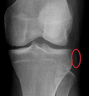 Segond Fracture Wikipedia
Posteromedial Corner Injury Of The Knee Radsource
Https Www Ajronline Org Doi Pdf 10 2214 Ajr 14 12693
 Lateral Collateral Ligament Lcl And Posterolateral Corner Plc Radiology Key
 Knee Pain Acute Trauma Diagnosis Imaging Part I El Paso Tx
 Mri Of The Posterolateral Corner Of The Knee Please Have A Look Sciencedirect
 Medial Capsular Ligament Knee Injury Radiology Case Radiopaedia Org
 Knee Capsule Radiology Reference Article Radiopaedia Org
 Magnetic Resonance Imaging In The Assessment Of Meniscal Anatomic Variants And Of The Perimeniscal Ligamentous Anatomy Potential Interpretation Pitfalls
 Hypermobile Lateral Meniscus Radsource
 Segond Fracture Radiology Reference Article Radiopaedia Org

Https Www Ajronline Org Doi Pdf 10 2214 Ajr 180 3 1800647
 Knee Joint Radiology Reference Article Radiopaedia Org
Http Www Ors Org Transactions 61 1693 Pdf
 Mri Of The Posterolateral Corner Injury A Concise Review Pacholke 2007 Journal Of Magnetic Resonance Imaging Wiley Online Library
 Normal Magnetic Resonance Imaging Anatomy Of The Capsular Ligamentous Supporting Structures Of The Knee Sciencedirect